Introduction
As we all know, heart problems are a serious issue . However, with early detection and proper treatment, many of these problems can be managed effectively. That’s why it’s so important to understand how heart problems are diagnosed and what options are available for treatment.
Over the next few slides, we’ll be discussing the most common heart complaints, the diagnostic tests used to identify them, how test results are interpreted, and the different treatment options available. By the end of this presentation, you’ll have a better understanding of how to protect your heart health and what steps you can take to ensure a healthy heart.
Common Heart Complaints
The most common heart complaints include chest pain, shortness of breath, irregular heartbeat, and fatigue. Chest pain is often described as a tightness or pressure in the chest that may radiate to the arms, neck, or jaw. Shortness of breath may occur during physical activity or at rest. An irregular heartbeat may feel like a fluttering or racing sensation in the chest. Fatigue is a feeling of tiredness or weakness that does not improve with rest.
Other common heart complaints include dizziness, lightheadedness, and swelling in the legs or ankles. Dizziness may be described as a feeling of being lightheaded or faint. Lightheadedness may be accompanied by a sensation of spinning or vertigo. Swelling in the legs or ankles may be a sign of fluid buildup caused by heart failure.
Diagnostic Tests

There are several diagnostic tests that can be used to identify heart problems.
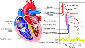
One of the most common tests is an electrocardiogram (ECG), which measures the electrical activity of the heart.
This test can help detect irregular heartbeats, heart attacks, and other heart problems.
During an ECG, electrodes are placed on the chest, arms, and legs, and the results are recorded on a graph.
- Echocardiogram: This is an ultrasound test that uses sound waves to create images of the heart’s structure and function. It can help assess the pumping function of the heart, identify valve abnormalities, measure the thickness of the heart walls, and evaluate blood flow.
- Stress test: Also known as an exercise stress test or treadmill test, this assesses the heart’s performance during physical activity. It involves walking on a treadmill or pedaling a stationary bike while being monitored for changes in heart rate, blood pressure, and ECG readings. It can help diagnose coronary artery disease and determine exercise tolerance.
- Cardiac catheterization: This invasive procedure involves inserting a thin tube (catheter) into a blood vessel, usually in the groin or arm, and advancing it to the heart. Contrast dye is injected, and X-ray images are taken to evaluate the coronary arteries, heart chambers, and valves. It can provide detailed information about blockages in the arteries and measure pressures within the heart.
- Cardiac MRI (Magnetic Resonance Imaging): This noninvasive test uses powerful magnets and radio waves to create detailed images of the heart. It provides information about the heart’s structure, blood flow, and function, making it useful in diagnosing heart diseases, such as heart muscle damage, congenital heart defects, and heart tumors.
- CT (Computed Tomography) Scan: This imaging technique uses X-rays and a computer to create cross-sectional images of the heart. It can help diagnose and assess conditions such as coronary artery disease, heart defects, calcium scoring for risk assessment, and pulmonary embolism.
- Holter monitor: It is a portable device worn by a patient that continuously records the heart’s electrical activity over a 24 to 48-hour period or longer. It can detect irregular heart rhythms that may not be captured during a short-term ECG.
- Blood tests: Blood tests can provide valuable information about heart health. These tests measure various markers such as cholesterol levels, blood sugar levels (for diabetes assessment), cardiac enzymes (for diagnosing a heart attack), and certain proteins associated with heart muscle damage or inflammation.
Another common test is an echocardiogram, which uses sound waves to create images of the heart. This test can help detect problems with the heart’s structure and function, such as valve problems or heart failure. During an echocardiogram, a technician will place a small device on the chest that emits sound waves and records the echoes that bounce back from the heart. The resulting images can provide valuable information about the heart’s health.
Interpreting Test Results
In an electrocardiogram (ECG), various waves, intervals, and segments are measured to evaluate the electrical activity of the heart. Here are the normal values for the main components of an ECG:
- P wave: The P wave represents atrial depolarization (contraction). Its duration should typically be less than 0.12 seconds.
- PR interval: The PR interval is measured from the beginning of the P wave to the beginning of the QRS complex. Its duration should typically be between 0.12 and 0.20 seconds.
- QRS complex: The QRS complex represents ventricular depolarization (contraction). Its duration should typically be less than 0.12 seconds.
- QT interval: The QT interval represents ventricular depolarization and repolarization. Its duration varies depending on the heart rate and should typically be corrected for heart rate using formulas such as the Bazett’s formula (QTc). The normal QTc interval is generally less than 0.44 seconds in men and less than 0.46 seconds in women.
- ST segment: The ST segment is measured from the end of the QRS complex to the beginning of the T wave. It should typically be at the same level as the baseline (isoelectric).
- T wave: The T wave represents ventricular repolarization. It should generally be upright in most leads and may vary in amplitude and shape.
- Heart rate: The normal resting heart rate for adults is usually between 60 and 100 beats per minute. However, heart rate can vary depending on factors such as age, physical fitness, and overall health.
It’s important to note that these values represent general guidelines, and interpretation of an ECG should consider the individual patient’s characteristics and clinical context. Any variations from the normal values may indicate underlying heart conditions and should be evaluated by a healthcare professional.
BLOOD ENZYMES
There are several blood enzymes that are commonly measured to assess heart health and detect possible damage to the heart muscle. Here are the normal ranges for some of the key cardiac enzymes:
- Troponin: Troponin is a highly sensitive and specific marker of heart muscle damage. The normal range for troponin levels is typically less than 0.04 ng/mL. Elevated levels of troponin may indicate a heart attack or other conditions that cause damage to the heart muscle.
- Creatine Kinase (CK): CK is an enzyme found in various tissues, including the heart muscle. However, it is not specific to the heart. There are three subtypes of CK: CK-MB, CK-MM, and CK-BB. CK-MB is the subtype that is most specific to the heart. The normal range for CK-MB is usually less than 6-7 ng/mL.
- CK-MB Index: The CK-MB index is the ratio of CK-MB to total CK. A CK-MB index greater than 2-3% is suggestive of heart muscle damage.
- Myoglobin: Myoglobin is a protein found in cardiac and skeletal muscle. Elevated myoglobin levels can be an early indicator of heart muscle damage. The normal range for myoglobin is typically less than 90-100 ng/mL.
THREAD MILL
During a treadmill exercise stress test, several parameters are monitored to assess the heart’s response to physical activity. Here are some normal values typically evaluated during a treadmill test:
- Heart Rate (HR): The target heart rate during exercise is generally calculated based on the individual’s age. It is often estimated using the formula: 220 minus age. For example, for a 40-year-old individual, the estimated maximum heart rate would be around 180 beats per minute (bpm). During the test, the heart rate should increase gradually as exercise intensity increases.
- Blood Pressure (BP): Blood pressure is monitored throughout the test, usually at regular intervals. The normal range for blood pressure during exercise varies depending on factors such as age, gender, and individual health. However, a general guideline is that systolic blood pressure (the top number) should increase with exercise, while diastolic blood pressure (the bottom number) should either remain stable or show a slight decrease.
- ECG Changes: Electrocardiogram (ECG) readings are continuously monitored during the test. Normal ECG changes during exercise include an increase in heart rate, consistent and regular heart rhythm, and appropriate ST segment response (elevation or depression within certain limits).
- Exercise Duration: The duration of the treadmill test can vary depending on the purpose and protocol used. Normal exercise duration for a standard treadmill test ranges from 6 to 15 minutes, depending on the individual’s fitness level.
It’s important to note that the interpretation of a treadmill test should consider the individual’s baseline health, exercise capacity, and presence of any underlying heart conditions. Abnormal findings during the test, such as significant chest pain, abnormal ECG changes, or abnormal blood pressure response, may indicate potential heart problems and should be further evaluated by a healthcare professional.
2D ECHO
A 2D echocardiogram, also known as a transthoracic echocardiogram (TTE), is a non-invasive imaging test that uses ultrasound to visualize the structure and function of the heart. Here are some normal values typically assessed during a 2D echo:
- Left ventricular ejection fraction (LVEF): LVEF represents the percentage of blood pumped out of the left ventricle with each heartbeat. The normal range for LVEF is typically 55% to 70%.
- Left ventricular end-diastolic diameter (LVEDD): LVEDD measures the size of the left ventricle at the end of diastole (relaxation phase). The normal range for LVEDD varies depending on factors such as age, gender, and body surface area. In adults, it is typically around 4.0 to 5.6 centimeters.
- Left ventricular end-systolic diameter (LVESD): LVESD measures the size of the left ventricle at the end of systole (contraction phase). The normal range for LVESD also varies depending on factors such as age, gender, and body surface area. In adults, it is usually around 2.2 to 4.0 centimeters.
- Left atrial diameter (LAD): LAD measures the size of the left atrium. The normal range for LAD varies depending on factors such as age, gender, and body surface area. In adults, it is typically around 2.5 to 4.0 centimeters.
- Interventricular septal thickness (IVS): IVS measures the thickness of the septum, which separates the left and right ventricles. The normal range for IVS is typically around 0.6 to 1.1 centimeters.
- Posterior wall thickness (PWT): PWT measures the thickness of the wall of the left ventricle. The normal range for PWT is typically around 0.6 to 1.1 centimeters.
- Valvular function: The 2D echo also assesses the function of heart valves, including the aortic valve, mitral valve, tricuspid valve, and pulmonic valve. The normal function of these valves includes proper opening and closure without significant regurgitation (backflow) or stenosis (narrowing).
It’s important to note that normal values can vary depending on factors such as age, gender, and individual patient characteristics.
HOLTER MONITOR
A Holter monitor is a portable device that records the electrical activity of the heart continuously over a 24 to 48-hour period or longer. It provides valuable information about the heart’s rhythm and can help detect any abnormalities. Here are some normal values typically considered when interpreting a Holter monitor:
- Heart Rate (HR): The normal resting heart rate for adults is usually between 60 and 100 beats per minute (bpm). During activities or periods of stress, the heart rate may increase, but it should return to the normal range during periods of rest.
- Heart Rhythm: A normal heart rhythm is typically characterized by a regular and consistent pattern of beats. It should not display significant irregularities or abnormal heart rhythms (arrhythmias) such as atrial fibrillation, ventricular tachycardia, or bradycardia.
- Premature Ventricular Contractions (PVCs): Occasional PVCs are common and usually not concerning. However, a high number of PVCs or specific patterns of PVCs may warrant further evaluation.
- ST Segment: The ST segment on the electrocardiogram (ECG) recorded by the Holter monitor should be relatively stable and close to the baseline. Significant ST segment elevation or depression may indicate myocardial ischemia or other cardiac conditions.
- T Wave: The T wave represents ventricular repolarization. It should generally be upright in most leads and may vary in amplitude and shape. Abnormal T wave inversions or flattening may indicate underlying heart problems.
- Symptoms: In addition to ECG findings, Holter monitors can help correlate symptoms such as chest pain, palpitations, dizziness, or shortness of breath with any abnormal heart rhythms or events captured during monitoring.
It’s important to note that the interpretation of Holter monitor results should be done by a healthcare professional experienced in reading and analyzing the data. They will consider the specific patient’s clinical history, symptoms, and other relevant factors when determining if any abnormalities are present.
It’s important to note that reference ranges may vary slightly between different laboratories and testing methods. Additionally, the interpretation of these enzyme levels should be done in conjunction with other clinical information and diagnostic tests to determine the underlying cause of heart complaints.
When it comes to interpreting test results for heart complaints, it’s important to consider a variety of factors. One of the most important things to look at is the overall pattern of the results, rather than just focusing on individual numbers. For example, an electrocardiogram (ECG) may show abnormalities in the heart rhythm or electrical activity, but it’s important to look at these findings in the context of the patient’s symptoms and medical history.
Another key factor in interpreting test results is understanding the normal ranges for different measurements. For example, a blood test may measure levels of certain enzymes or proteins that can indicate heart damage, but the significance of these levels depends on the patient’s age, sex, and other factors. By looking at all of the available information and considering each piece in context, healthcare providers can make more accurate diagnoses and develop effective treatment plans.
Treatment Options
Medication is often the first line of treatment for heart problems. Depending on the type of heart problem, patients may be prescribed medications such as beta-blockers, ACE inhibitors, or diuretics. These medications work by reducing blood pressure, slowing the heart rate, and improving blood flow to the heart. While these medications can be effective in controlling symptoms and preventing further damage to the heart, they do come with side effects that need to be carefully monitored.
Lifestyle changes can also play a crucial role in treating heart problems. Patients may be advised to make changes such as quitting smoking, eating a healthy diet, exercising regularly, and managing stress. These changes can help improve overall heart health and reduce the risk of further heart problems. However, making these changes can be difficult and require a lot of effort and commitment from the patient.
Surgery may be necessary for more serious heart problems, such as blocked arteries or damaged heart valves. Procedures such as angioplasty, bypass surgery, or valve replacement can help improve blood flow to the heart and reduce symptoms. While these procedures can be very effective, they also carry significant risks and require a longer recovery time than other treatment options.
Conclusion
In conclusion, we have discussed the most common heart complaints and their symptoms, as well as the different diagnostic tests that can be used to identify heart problems. We also explored how test results are interpreted and what they can reveal about a patient’s heart health. Finally, we looked at the different treatment options for heart problems, including medication, lifestyle changes, and surgery.
It is important to remember that early detection and treatment of heart problems can greatly improve outcomes and quality of life. By taking steps to protect your heart health, such as getting regular check-ups and making healthy lifestyle choices, you can reduce your risk of developing heart problems. Remember, your heart health is in your hands!


